The project aims at investigating new signal reconstruction methods from diffusion magnetic resonance imaging (dMRI) allowing an improved description of microstructural properties and at assessing their impact on the accurate anchoring of the transcranial magnetic stimulation (TMS) electromagnetic field by the consequentially identified neuronal axons’ orientation and microstructural features.
Realistic modeling of TMS brain stimulation requires the integration of microstructural properties with modeling of the electromagnetic field pulse propagation in complex (non homogeneous) structures.
The most appropriate way for tackling this problem is to inject microstructural models into dynamic electro-magnetic (EM) propagation models and validating them in real operational conditions. Though, this is a very difficult problem for which a solution is far from being reached in the state-of-the-art (SOA).
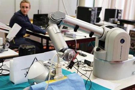
In this project, we focus on two specific issues intervening in the global solution: modeling of the diffusion signal and assessment of the impact of the devised solution on the characterization of neuronal targeting in TMS, using diffusion tensor imaging (DTI) for benchmarking. The validation will be performed by multi-modality assessment by two ad-hoc experiments. These will be performed using the neuronavigation system (NSS) and the Video-EEG facilities that are available at the Navigation Lab (NAVLab), complemented by a 32-channel EEG cap enabling improved spatial resolution.


